This is a slightly invasive procedure for the treatment of painful vertebral compression fractures, which are fractures relating the vertebral bodies that make up the spinal column. At the point when a vertebral body fractures, the typical rectangular shape of the bone ends up compressed, as a result, triggers the pain.
These compression fractures may include the breakdown of one or more vertebrae in the spine and are a common aftereffect of osteoporosis. Osteoporosis is a disorder that outcomes in a loss of normal bone density, mass and strength, driving towards a condition in which bones turn out to be more and more porous, and helpless against breaking easily.
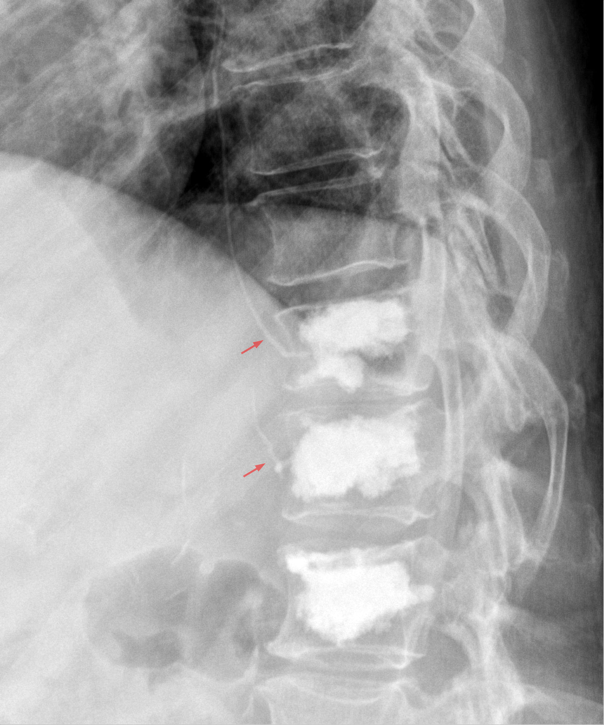
In order to perform kyphoplasty treatment, doctors utilize image guidance, in most cases fluoroscopy, in order to inject a blend of cement into the cracked bone through a hollow needle.
Kyphoplasty Treatment Before and During the Procedure
Preparation for the Procedure:
Based on the fact that kyphoplasty treatment is a surgical procedure, the doctor will most likely require some blood tests prior to the day of the procedure. Furthermore, X-Ray or MRI will enable the doctor to see the area or areas that need repair.
In the process of preparation, an intravenous line will be set in a vein in order to deliver anesthesia. You may likewise get pain and anti-nausea medications, as well as antibiotics, to avoid infection. The patient most likely additionally be connected to the heart, pulse, and blood pressure monitors.
Kyphoplasty Treatment Procedure:
In these sort of procedures, patients are required to lie down on the stomach. The area in which the needle will be inserted is shaved on the off chance that obligatory and afterwards cleaned and sterilized.
Following Are The Steps Performed By The Surgeon:
- A hollow needle is inserted into the skin with the guidance of fluoroscopy, this is a kind of X-Ray. They guide the needle through the muscles and into the right position in the bone.
- In the next step, an inflatable balloon is inserted into the trocar.
- After that, the balloon is inflated in order to make the space required for the bone cement.
- As soon as space has opened up, the blend is injected in order to fill it. In this case, imaging tests work in a way to assist the surgeon to confirm that the blend is distributed appropriately.
- As soon as the mixture is set up, the needle is taken out.
- After that, the area is bandaged. In this case, stitches won’t be required.
- IV and monitoring equipment are removed.