Welcome to Pain Management Institute

To make an appointment for any of our locations, please call
  815.412.6166

All posts by admin

2019-04-02

What Is Epidural Lysis And How The Procedure Is Done?

The condition is commonly caused by leakage into the lumbar spine’s epidural space after the surgical process. Adhesions can result in constant leg pain, yet doctors are skilled for breaking up the scar tissue that works in a way to cause pain with the utilization of a particular catheter and blend of medications. This is actually an epidural lysis procedure.

In most cases, epidural adhesions happen after spine surgery, however, in some cases, this can likewise happen after disc herniation because of leakage of disc material into epidural space. The objective of epidural lysis procedure is to precisely and chemically break off unnecessary scar tissue in the epidural space in order to diminish nerve disturbance and potentially ease the pain.

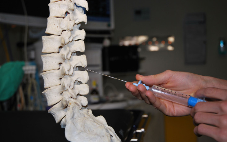
How Is The Epidural Lysis Procedure Done?

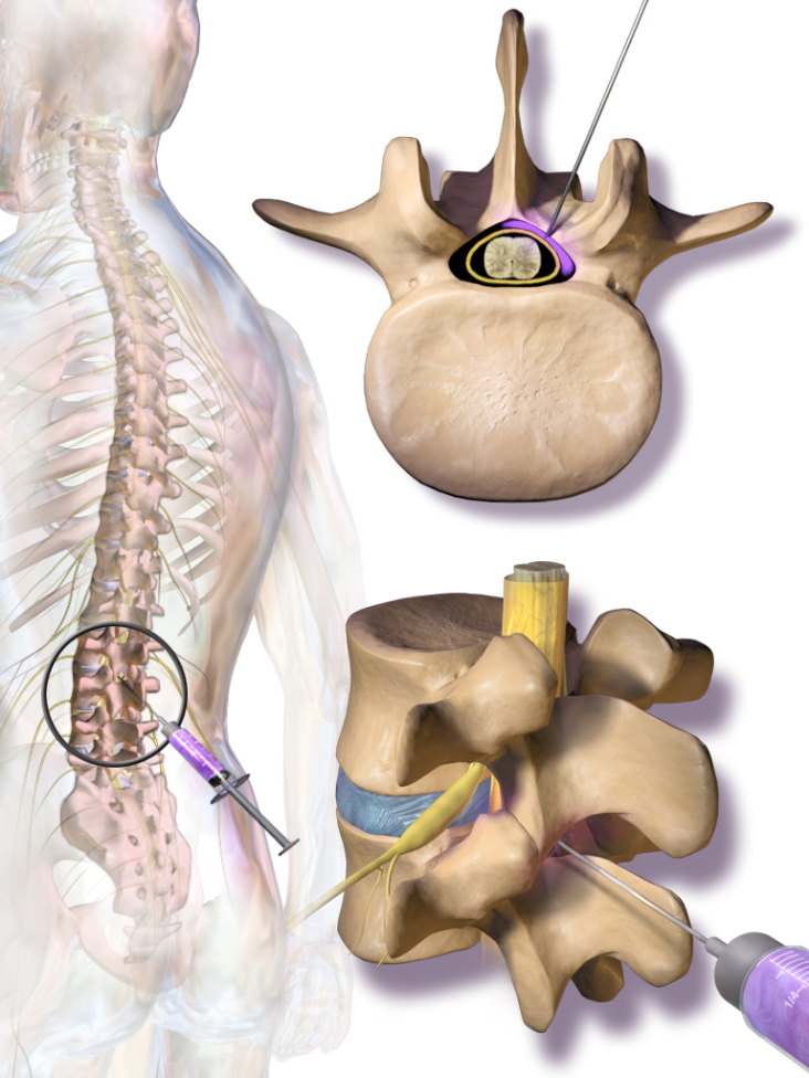
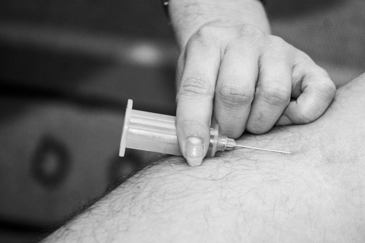
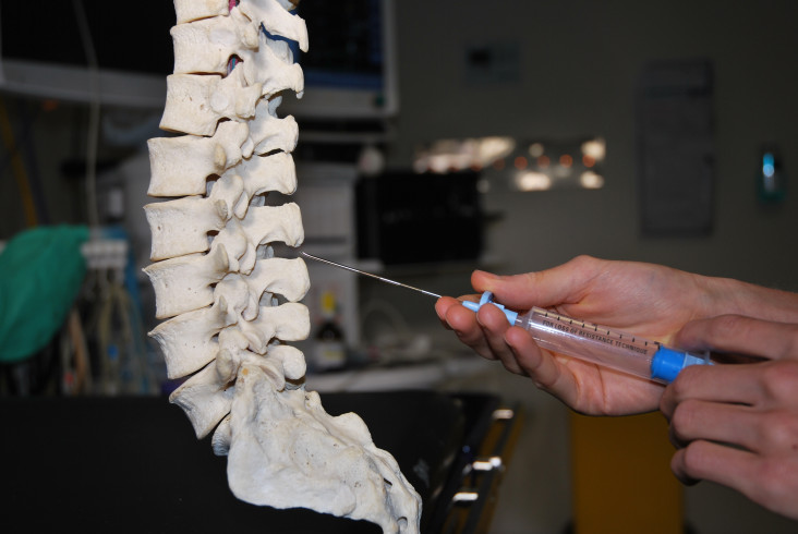
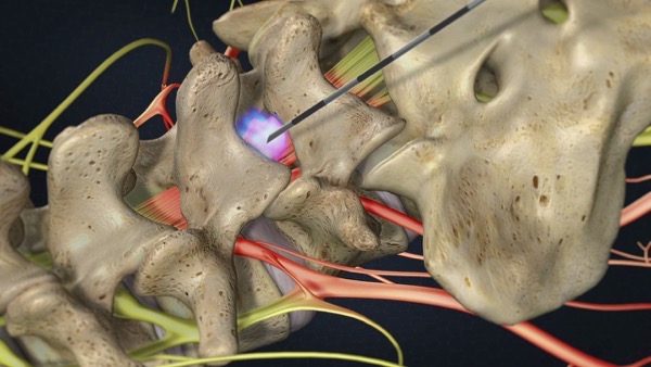
The patient will be required to lie on the stomach, then the monitors will be attached in order to screen the heart rate, blood pressure, and oxygenation. After the patient has been sedated, the back will be brushed with an antiseptic solution and drape the back with sterile material. The doctor will numb the skin with a local anesthetic. After that, the doctor will insert a particular needle through the skin simply over the tailbone region into the epidural space.

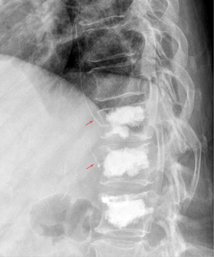
An X-Ray dye will be injected into the needle to picture the scarred region. A particular catheter will be inserted through the needle to the scarred zone in order to break the adhesions. Furthermore, different liquids will be injected through the catheter with an objective to diminish the pain, break down scar tissue, and decrease inflammation. Regular medications utilized are steroids, local anesthetic, enzyme and hypertonic saline.

Suitable Candidates for Epidural Lysis Procedure

In most cases, the procedure is utilized with patients who have constant lower back and leg pain because of epidural adhesions after lumbar (lower) spine surgery or an injury. However, symptoms include lower back pain (normally a consistent, burning pain), radicular pain, (for example, sciatica which shoots from the spine into the thighs, calves or feet), muscle spasms, motor/sensory or reflex changes, and restricted flexibility in the thighs and calves.

Besides treating unnecessary scarring, this procedure can be utilized to treat a scope of other back and spine conditions, including:

  • Herniated or prolapsed intervertebral discs (particularly when not surgically correctable).
  • Metastatic carcinoma of the spine (driving towards compression fracture).
  • Degenerative spondylosis (influencing numerous levels of the spine).
  • Spinal (back) stenosis (a reduction of the open spaces within the spine).
  • Cervical (neck) stenosis.
  • Post-laminectomy syndrome (failed back surgery).
  • Degenerative arthritis of the spine.
  • Occipital neuralgia.

Epidural lysis procedure is certainly not a “first line” treatment. However, in most cases, patients who go through this treatment have attempted more conservative treatment alternatives, (for example, medications and epidural steroid injections), which have been unsuccessful to ease the pain.

Read More
Blausen_0354_EpiduralSteroidInjection

What Is An Epidural Steroid Injection And Who Is A Suitable Candidate?

This is a common procedure for treating inflammation related to low back related leg pain, or neck related arm pain. In both of these conditions, the spinal nerves turn out to be inflamed because of tightening of the passages where the nerves travel as they go down or out of the spine.

Why There Is A Need Of Epidural Steroid Injections?

Tightening of the spinal sections can result from a range of causes, this includes disc herniation, bone spurs, thickening of the ligaments in the spine, joint cysts, or even unusual arrangement of the vertebrae. The epidural space is a fat filled that involves the spinal sac and gives cushioning to the nerves and spinal cord.

In this case, steroids put into the epidural space have a strong anti-inflammatory response that works in a way to diminish pain and enable patients to improve mobility. In spite of the fact that steroids don’t change the fundamental condition, they can work in a way to break the cycle of pain and inflammation and enable the body to compensate for the condition. As a result, the epidural steroid injections can give benefits that outlast the impacts of the steroid itself.

The Suitable Candidates for Epidural Steroid Injections

Individuals suffering from pain in the neck, arm, low back, or leg may turn out to be suitable candidates for epidural steroid injections. In particular, those with the following conditions:

Spinal Stenosis:

A tightening of the spinal canal and nerve root canal can result in back and leg pain, particularly when walking.

Spondylolisthesis:

A weakness or fracture in the middle of the upper and lower facets of a vertebra. In the event that the vertebra slips forward, it can compress the nerve roots resulting in pain.

Herniated Disc:

The gel-like material inside the disc can result in bulging or rupture through a weak zone around the wall. Irritation, pain, and swelling happen when this material crushes out and interacts with a spinal nerve.

Degenerative Disc:

A breakdown or aging of the intervertebral disc resulting in the breakdown of the disc space, tears in the annulus, and growth of bone spurs.

Sciatica:

Pain that progresses along the sciatic nerve in the buttocks and down the legs. It is normally resulted by compression of the fifth lumbar or first sacral spinal nerve.

The utilization of epidural steroid injections has demonstrated positive outcomes for certain patients in the treatment of painful inflammatory conditions. The injections can likewise help decide if surgery may be advantageous for pain related to a herniated disc. At the point when symptoms meddle with rehabilitative activities, epidurals can work in a way to ease the pain enough so patients can proceed with their physical therapy.

Read More
photo-1513315849661-23219170a967

What Is A Facet Block Injection? The Procedure and Purpose of Facet Block Injection

This is a treatment that gives brief relief from back pain, just as a test to see if facet joints are the source of a patient’s back pain. In this case, the injection is inserted into the facet joints, which are situated on each side of the vertebra and associate the vertebra of the back together. The injection works in a way to diminish inflammation in the tissue of the facet joint.

The facet block injection is performed with requiring the patient lying on the abdomen. Preceding the injection, the site of the injection is cleaned and numbed with local anesthetic. The medication is inserted with the help of X-Ray guidance.

Prior to the Facet Block Injection

The patient will be asked to provide information in regards to the ongoing issues, past medical history, and medications. The patient will be guided with complete guidelines on the best way to plan for the coming procedure. It is advisable to carry a recent MRI with you. The patient will likewise be asked to hold any blood thinning medications, for example, Coumadin, Plavix, Aspirin, and any NSAIDS.

After The Facet Block Injection

After the procedure, you may have brief numbness or faintness of your extremities enduring up to 6 hours. In some cases, it might be problematic to walk because of an absence of sensation in your foot. As this decreases, a few patients report expanded inconvenience. This can be linked with disturbance from the steroid and will resolve. You should start to observe relief from pain after 3-7 days of the procedure. However, there is no certainty how much relief from pain you will get. Fruitful outcomes rely upon your diagnosis.

What Purposes Does A Facet Block Injection Serve?

This treatment serves two needs: diagnosis and treatment. On the off chance that the patient gets 50% or more noteworthy relief from pain like 2 hours after the procedure, it can be anticipated the facet joint is a source of the pain in this case, as may occur with joint pain or injury. Once the diagnosis is confirmed with repeat injections, the facet block is then used to diminish pain using corticosteroids.

How Soon Can You Resume Everyday Activities After The Procedure Of Facet Block Injection?

When allowed to go home, you will be advised to rest for 24 hours and avoid driving or operating machinery for 24 hours. A sum of three injections inside a six-month timespan can be performed. In this case, injections must be spaced at least two weeks apart and you should contact the doctor in the middle of injections to get a new prescription for each.

Read More
Vertebre-IMG_0032

What Is Intradiscal Electrothermal Therapy And How Does It Work?

This is a fairly new technique that is slightly invasive. It is performed in cases when patients experience chronic discogenic back pain. It is believed that the discogenic back pain takes place due to the intervertebral disc degeneration. As indicated by research, discogenic pain occurs somewhere in the range of 28% and 43% of back pain cases in the United States. Moreover, additional researches have demonstrated that discogenic pain is one of the main reasons for low back pain in the United States.

Numerous patients going through discogenic back pain respond to traditional treatment alternatives including non-steroidal anti-inflammatory drugs (NSAIDs), exercise, and physical therapy. On the other hand, there are a few patients who fail to respond to these treatment strategies. Unfortunately, there are very few treatments accessible for patients amongst these traditional techniques and surgery. Nonetheless, intradiscal electrothermal therapy is a comparative traditionalist treatment strategy that can be offered to patients rather than surgical procedure.

How Does Intradiscal Electrothermal Therapy Work?

As we age or due to an injury, the tears or cracks occur in the walls of the intervertebral disc. These tears can end up filled with small nerve endings and blood vessels, however, in some cases, this turns out to be chronic pain. In principle, the intradiscal electrothermal therapy works in a way to thicken and strengthen the disc wall by applying controlled heat to the disc wall.

To begin with the procedure, a small tube known as a catheter is inserted into the disc with fluoroscopic direction. The catheter is then moved to accomplish a 360-degree entrance. Heat is brought into the disc by means of the catheter, steadily advancing around 150° F to roughly 200° F. The whole intradiscal electrothermal therapy is performed on an outpatient premise and just requires around 60 minutes, with an extra hour of recuperation time.

A few patients state quick relief from discomfort, however, all things considered, noteworthy relief and improvement in function happen as the disc heals. This procedure, for the most part, takes four to twelve weeks. Patients usually come back to a basic routine within one week of the procedure. After around six weeks, progressively strenuous physical activity, in combination with a formal spinal- strengthening program, may start.

Conclusively, intradiscal electrothermal therapy is a generally conservative treatment alternative that is accessible to patients who go through discogenic pain that has been unsuccessful to respond to customary treatment techniques. Different research studies have demonstrated the safety and adequacy of this procedure and have demonstrated positive outcomes for the usage of this technique in patients experiencing discogenic pain.

Read More
Intercostal-Nerve-Block

Intercostal Nerve Block Procedure, Anatomy and Benefits

This procedure is utilized to deal with pain in the chest and upper abdomen areas. The intercostal nerve is situated between ribs all through the thoracic area. In order to ease inflammation of these nerves, the anesthetics, steroids, or other medications are injected as they inhibit the transmission of pain signals. The procedure of the intercostal nerve block may likewise be utilized to diagnose pain and distinguish the direction of the pain signal. Following conditions can be treated with this procedure:
• Neuropathic pain in the chest related to herpes zoster or shingles
• Postoperative pain of chest or upper abdominal surgery
• Chronic pain after mastectomy
• Rib fracture pain
• Pain associated with the formation of scar tissue after surgery
• Somatic pain secondary to metastatic cancer to the ribs
The intercostal nerve conveys pain signals from the muscles, bones, and skin to the brain, and a block can be successful in easing pain without the adverse effects of opioid pain medications. It is easy for the physician to access the nerve. A single intercostal nerve block may result in permanent help with discomfort.

Anatomy of Intercostal Nerve Block

The intercostal nerves begin from 12 paired thoracic nerve roots that are closely connected with the thoracic ribs. As the thoracic nerve roots emerge from the intervertebral foramen, they quickly split into the ventral rami that structure the intercostal nerves and the posterior rami. Anterior branches structure the gray and white rami communicantes of the thoracic sympathetic chain. The posterior rami innervate the zygapophyseal joints, muscles and skin of the thoracic midline and paraspinous region of the back.
The lateral cutaneous nerve splits into the back and anterior branches that innervate the skin of the chest wall from the scapular line to the midclavicular line. The intercostal nerve proceeds anteriorly inside the costal groove between the internal intercostal muscle layer and the external area of the innermost intercostal muscle, yet as it advances anteriorly, it once more emerges internally to the deepest intercostal muscle.

Advantages of Intercostal Nerve Block

This is a short outpatient procedure that is suitable for easing the pain of the muscles, bones, and skin in the chest and upper abdomen. Patients having hard to treat neuropathic or metastatic bone cancer pain may observe an intercostal nerve block lessens their pain or get rid of it. The intercostal nerve block is a decent alternative to opioid pain medications, which convey adverse effects the patient will most likely be unable to endure. On the off chance that the procedure is effective, the patient is a suitable candidate for repeat injections to treat repeating pain.

Read More
320px-Innermost_intercostal_muscles_back

Intercostal Nerve Block Procedure, Anatomy and Benefits

This procedure is utilized to deal with pain in the chest and upper abdomen areas. The intercostal nerve is situated between ribs all through the thoracic area. In order to ease inflammation of these nerves, the anesthetics, steroids, or other medications are injected as they inhibit the transmission of pain signals. The procedure of the intercostal nerve block may likewise be utilized to diagnose pain and distinguish the direction of the pain signal. Following conditions can be treated with this procedure:

  • Neuropathic pain in the chest related to herpes zoster or shingles
  • Postoperative pain of chest or upper abdominal surgery
  • Chronic pain after mastectomy
  • Rib fracture pain
  • Pain associated with the formation of scar tissue after surgery
  • Somatic pain secondary to metastatic cancer to the ribs

The intercostal nerve conveys pain signals from the muscles, bones, and skin to the brain, and a block can be successful in easing pain without the adverse effects of opioid pain medications. It is easy for the physician to access the nerve. A single intercostal nerve block may result in permanent help with discomfort.

Anatomy of Intercostal Nerve Block

The intercostal nerves begin from 12 paired thoracic nerve roots that are closely connected with the thoracic ribs. As the thoracic nerve roots emerge from the intervertebral foramen, they quickly split into the ventral rami that structure the intercostal nerves and the posterior rami. Anterior branches structure the gray and white rami communicantes of the thoracic sympathetic chain. The posterior rami innervate the zygapophyseal joints, muscles and skin of the thoracic midline and paraspinous region of the back.

The lateral cutaneous nerve splits into the back and anterior branches that innervate the skin of the chest wall from the scapular line to the midclavicular line. The intercostal nerve proceeds anteriorly inside the costal groove between the internal intercostal muscle layer and the external area of the innermost intercostal muscle, yet as it advances anteriorly, it once more emerges internally to the deepest intercostal muscle.

Advantages of Intercostal Nerve Block

This is a short outpatient procedure that is suitable for easing the pain of the muscles, bones, and skin in the chest and upper abdomen. Patients having hard to treat neuropathic or metastatic bone cancer pain may observe an intercostal nerve block lessens their pain or get rid of it.  The intercostal nerve block is a decent alternative to opioid pain medications, which convey adverse effects the patient will most likely be unable to endure. On the off chance that the procedure is effective, the patient is a suitable candidate for repeat injections to treat repeating pain.

Read More
Epidural_needle_insertion_between_the_spinous_processes_of_the_lumbar_vertebrae

Intrathecal Pump Therapy Procedure and Possible Complications and Who Are the Suitable Candidates for Intrathecal Pump Therapy

This is a technique for delivering medication straight to the spinal cord. In case, all other traditional techniques have been unsuccessful to relieve long term symptoms, then a pain pump might be an alternative treatment. Based on the fact that the medication is delivered straight to the spinal cord, it can work in a way to control the symptoms with a much smaller dose than is required with oral medication. The objective of intrathecal pump therapy is to efficiently control the symptoms and to diminish oral medications.

The Anticipated Outcomes after the Procedure

The patient will wake up in the postoperative recuperation facility. Furthermore, the patient’s blood pressure, heart rate, and respirations will be observed, and the pain will be addressed. In most cases, the patient is allowed to go home the same day. The patient will be provided with the written instructions to follow. Outcomes will differ contingent upon the underlying condition being dealt with and its seriousness.

Possible Complications after Intrathecal Pump Therapy

In this case, the adverse effects are minimal, in spite of the fact that they do exist. Similarly, as with all surgeries, complications may incorporate infection and bleeding. The catheter could move or end up blocked, or the pump could quit working (however, rare). The fluid can gather around the pump resulting in a clear watery discharge from incisions or result in a headache. However, these normally vanish on their own but may require a drain.

Adverse effects from the medications may incorporate:

  • Respiratory Depression
  • Twitching
  • Muscle Spasm
  • Urinary Retention
  • Constipation
  • Nausea
  • Vomiting
  • Dizziness
  • Anxiety
  • Depression
  • Edema

Contingent upon how much medication the pump delivers, the battery will eventually be required to replace.

Possible Limitations after Intrathecal Pump Therapy

The patient will be advised to stay away from particular activities for around six to eight weeks in order to avoid movement of the catheter. Moreover, no bending, twisting, stretching, raising the arms over the head or lifting heavy objects. Prevent sleeping on stomach. No climbing stairs or sitting for extensive periods.

The patient will be advised to avoid driving for 2 to 4 weeks after surgery or until examined with their surgeon. Furthermore, housework and yard-work are not allowed until the first follow-up office visit. This incorporates gardening, mowing, vacuuming, ironing, and loading/unloading dishwasher, or dryer.

Who Are The Suitable Candidates?

This procedure incorporates individuals for whom traditional treatments have been unsuccessful. The decision of treatment relies upon the sort of pain, how serious it is, and how you react to pain treatment. In the event that the doctor thinks you are a decent candidate for intrathecal pump therapy, you can go through a trial screening test so you can go through the treatment to check whether you are a decent candidate.

Read More
Zementaustritt_nach_Kyphoplastie_77jw_-_Roe_-_001

What Is Kyphoplasty And How The Procedure Is Performed?

This is a slightly invasive procedure for the treatment of painful vertebral compression fractures, which are fractures relating the vertebral bodies that make up the spinal column. At the point when a vertebral body fractures, the typical rectangular shape of the bone ends up compressed, as a result, triggers the pain.

These compression fractures may include the breakdown of one or more vertebrae in the spine and are a common aftereffect of osteoporosis. Osteoporosis is a disorder that outcomes in a loss of normal bone density, mass and strength, driving towards a condition in which bones turn out to be more and more porous, and helpless against breaking easily.

In order to perform kyphoplasty treatment, doctors utilize image guidance, in most cases fluoroscopy, in order to inject a blend of cement into the cracked bone through a hollow needle.

Kyphoplasty Treatment Before and During the Procedure

Preparation for the Procedure:

Based on the fact that kyphoplasty treatment is a surgical procedure, the doctor will most likely require some blood tests prior to the day of the procedure. Furthermore, X-Ray or MRI will enable the doctor to see the area or areas that need repair.

In the process of preparation, an intravenous line will be set in a vein in order to deliver anesthesia. You may likewise get pain and anti-nausea medications, as well as antibiotics,  to avoid infection. The patient most likely additionally be connected to the heart, pulse, and blood pressure monitors.

Kyphoplasty Treatment Procedure:

In these sort of procedures, patients are required to lie down on the stomach. The area in which the needle will be inserted is shaved on the off chance that obligatory and afterwards cleaned and sterilized.

Following Are The Steps Performed By The Surgeon:

  • A hollow needle is inserted into the skin with the guidance of fluoroscopy, this is a kind of X-Ray. They guide the needle through the muscles and into the right position in the bone.
  • In the next step, an inflatable balloon is inserted into the trocar.
  • After that, the balloon is inflated in order to make the space required for the bone cement.
  • As soon as space has opened up, the blend is injected in order to fill it. In this case, imaging tests work in a way to assist the surgeon to confirm that the blend is distributed appropriately.
  • As soon as the mixture is set up, the needle is taken out.
  • After that, the area is bandaged. In this case, stitches won’t be required.
  • IV and monitoring equipment are removed.


Read More
Spinal_Decompression

Microdiscectomy Spine Surgery’s Purpose of Procedure and Success Rate

In the case of microdiscectomy spine surgery, an exceptionally specialized procedure is performed on the spine, yet it is essentially utilized to decrease pain in another part of the body altogether. It is a fact that the spine is consists of a chain of bones each cushioned by an elastic disc that supports in both shock absorption and flexibility. On the off chance that a disc in the lower back turns out to be herniated and as a result, pushes on a spinal nerve, consequently, pain can radiate down a patient’s leg.

The procedure of microdiscectomy spine surgery is slightly invasive executed on the spine that works in a way to help most individuals feel prompt relief and return home the same or next day. The procedure is executed through a small incision, generally, one to one and-a-half – inches length, the objective of the procedure is to relieve the pressure on the affected spinal nerve by taking out a part of the bone or disc resulting in the pressure and pain.

During the microdiscectomy spine surgery, the spine’s structure remains intact, in light of the fact that this phenomenal surgery doesn’t require the cutting of joints, ligaments, or muscles. Patients who go through the procedure return to their normal life earlier, without leg pain, offering improved comfort and quality of life.

The Purpose of Microdiscectomy Spine Surgery

The key motive of this procedure is to surgically take out a part of the disc that is pushing on the nerve and hence, causing the pain. The objective is to take the pressure off the nerve yet at the same time retain as much of the disc as could be possible. The whole disc isn’t taken out, only the herniated part. The process usually takes on an average l around 60 minutes.

A timely return to normal routine is depicted with three exceptions. Heavy lifting, bending from the waist and delayed sitting are discouraged for the initial six weeks after surgery. Most patients can go up and down stairs when they return home from the hospital.

Success Rate of Microdiscectomy Spine Surgery

As per the statistics, the success rate of the procedure is commonly high, one extensive medical study presenting good or astounding outcomes generally for 84% of individuals who have gone through the strategy.

The report additionally indicates a few advantages for surgery, when contrasted with nonsurgical treatment, however, the distinction diminishes after some time in specific cases. One vast examination found that individuals who had surgery for a lumbar herniated disc had more noteworthy improvement in symptoms for as long as two years than the individuals who did not have surgery.

Read More
lumepisteroid_1280

Racz Epidural Neurolysis Benefits and Suitable Candidate

In the case of RACZ epidural neurolysis, a series of injections or an injection utilized in the area of the spine. This method is utilized as a treatment for chronic back pain. The repeated injections are utilized four times during the two days in house treatment. It is a fact that the back pain most of the times is complex and has numerous causes. Going through interventional pain treatment for chronic back pain has demonstrated that the pain frequently takes place because of scar tissue that is either from injury, disc herniation, or past back surgical procedure.

In most cases, RACZ epidural neurolysis can work in a way to avoid the requirement for spinal surgery with chronic back pain and sciatic pain. Based on the fact that the treatment can possibly give prompt and vital long term ease from chronic pain and uneasiness in the lumbar spine, without the requirement for spinal surgery. Furthermore, this treatment is also successful in easing lower back pain and transmitting sciatic pain.

How Can The Doctor Determine, In Case, The Racz Epidural Neurolysis Treatment Is Feasible For You?

The treatment is only suggested in conditions of extreme, chronic pain. Patients are required to have somewhere around three months history of unsuccessful, conventional treatment, for back or sciatic pain in order to be a suitable candidate for RACZ epidural neurolysis. In most conditions, RACZ epidural neurolysis can likewise turn out to be effective after a successful micro-surgical nerve decompression, as this surgical procedure can leave the spine with agonizing surgical scarring, or annoyance in the region of the spinal cord.

Nerve root irritation can be determined through MRI-imaging. Around six weeks ought to be allowed in the middle of the recent epidural injection and the RACZ catheter for security reasons.

Advantages of Racz Epidural Neurolysis

The procedure totally maintains a strategic distance from tissue injury. The procedure utilizes the natural openings in the spine so as to get to the epidural space. Thus it turns out to be the least invasive type of surgical pain therapy accessible. The treatment is mainly useful after unsatisfactory outcomes from micro-surgical treatment (open spine surgery). Contingent upon conditions, up to 50% of patients feel no adequate relief from pain following micro-surgery, despite the fact that the essential issue ought to have been addressed.

The zone around the spine is especially weak for surgical procedures because of its high density of nerve tissue. Scar formation after open spinal surgery can potentially drive towards chronic irritation and loss of function, in this way making a secondary issue.

Read More